Categories
3D Printing AR / VR Digital Healthcare HoloLens Image Processing Windows

Visualizing MRI & CT Scans in Mixed Reality / VR / AR, Part 3: 3D Model Maker

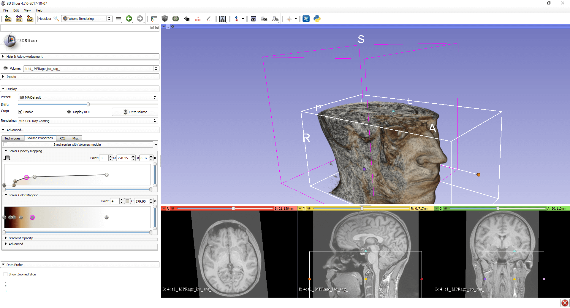
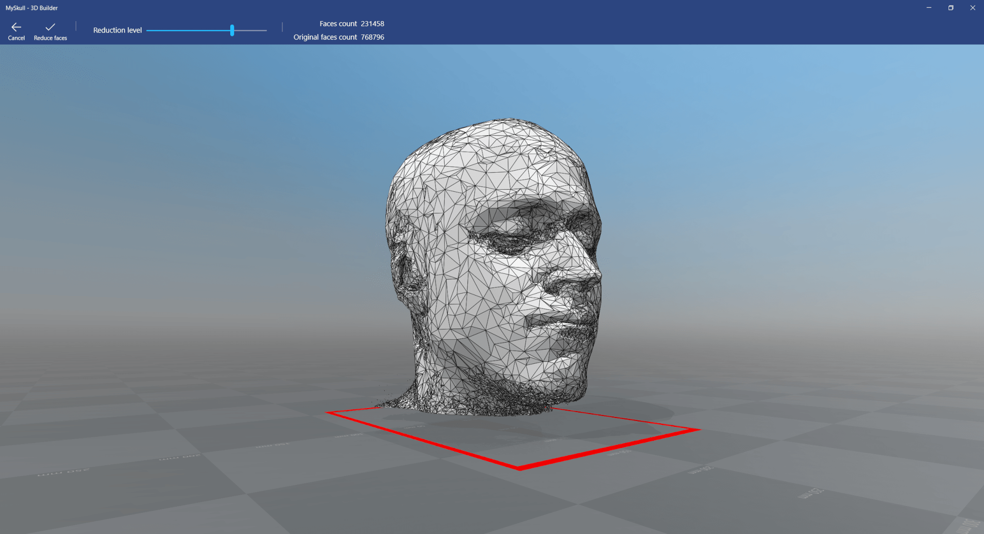
So far, we’ve created a volume rendering of an MRI / CT / Ultrasound scan. This is based on Voxels. For 3D printing and highly performant visualization in AR / VR scenarios, we need to create and export a polygon-based model. For the first step, we will use the Grayscale Model Maker and export the 3D Model as .stl to further prepare the model.

To create a 3D model, we have two main options in 3D Slicer:

  • Grayscale Model Maker: directly uses grayscale values from the image data. A threshold defines the surfaces. The model maker also takes care of smoothing the surfaces and reducing the polygon count.
  • Model Maker: this requires labels or discrete data to build a 3D model, meaning you have to segment the image data.

As a first step, we will use the Grayscale Model Maker, and later explore the more advanced options offered by segmentation and the Model Maker.

Categories
3D Printing AR / VR Digital Healthcare HoloLens Image Processing Windows

Visualizing MRI & CT Scans in Mixed Reality / VR / AR, Part 2: 3D Volume Rendering

After importing the MRI / CT / Ultrasound data into 3D Slicer in part 1, we’re ready for the first 3D visualization inside the medical software through 3D Volume Rendering. This is a major step to export the 3D model to Unity for visualization through Google ARCore or Microsoft HoloLens, or for 3D printing.

Slices in 3D View

After optimizing brightness and contrast of the image data, the easiest way of showing the data in 3D is to visualize the three visible slices (planes: axial / top / red; sagittal / side / yellow; coronal / frontal / green view) in the 3D view. This gives a good overview of the position and the relation of the slices to each other.

Categories
3D Printing AR / VR Digital Healthcare HoloLens Image Processing Windows

Visualizing MRI & CT Scans in Mixed Reality / VR / AR, Part 1: Importing Data

Some of the best showcases of Mixed Reality / VR / AR include 3D visualizations of MRI (magnetic resonance imaging), CT (computer tomography) or ultrasound scans. 3D brings tremendous advantages for analyzing the scanned images compared to only viewing 2D slices. Additionally, a good visualization brings value to patients who can gain a better understanding if they can easily explore their own body.

As part of the 3D information visualization lecture at the FH St. Pölten, I’m giving an overview of the process of converting an MRI / CT / ultrasound scan into a hologram that you can view on the Microsoft HoloLens or with Google ARCore. This blog post series explains the hands-on parts, so that you can easily re-create the same results using freely available tools.

Categories
Image Processing

20 – 30% Better JPEG compression from Google? My Test Results.

The blog post from Google sounds intriguing – 20% to 30% better JPEG image compression at the same visual quality through the Guetzli encoder. That has potential for a huge speed increase of websites.

While there are of course a lot of other better image formats around than JPEG (e.g., WebP or JPEG 2000), time has shown that it’s more or less impossible for them to gain any traction. It’s a pity, but on the other hand ensures that pretty much every device available on the market right now can load and show all websites.

Testing Guetzli!

To check the new Guetzli JPEG encoder out myself, I downloaded the binary executable of v1.0 and put it through some very quick tests.